Nervous Tissue · Anatomy and Physiology
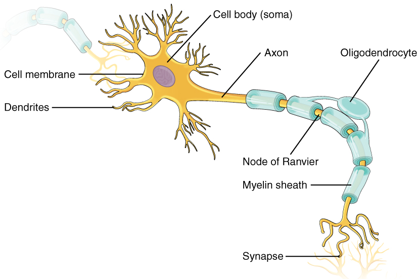
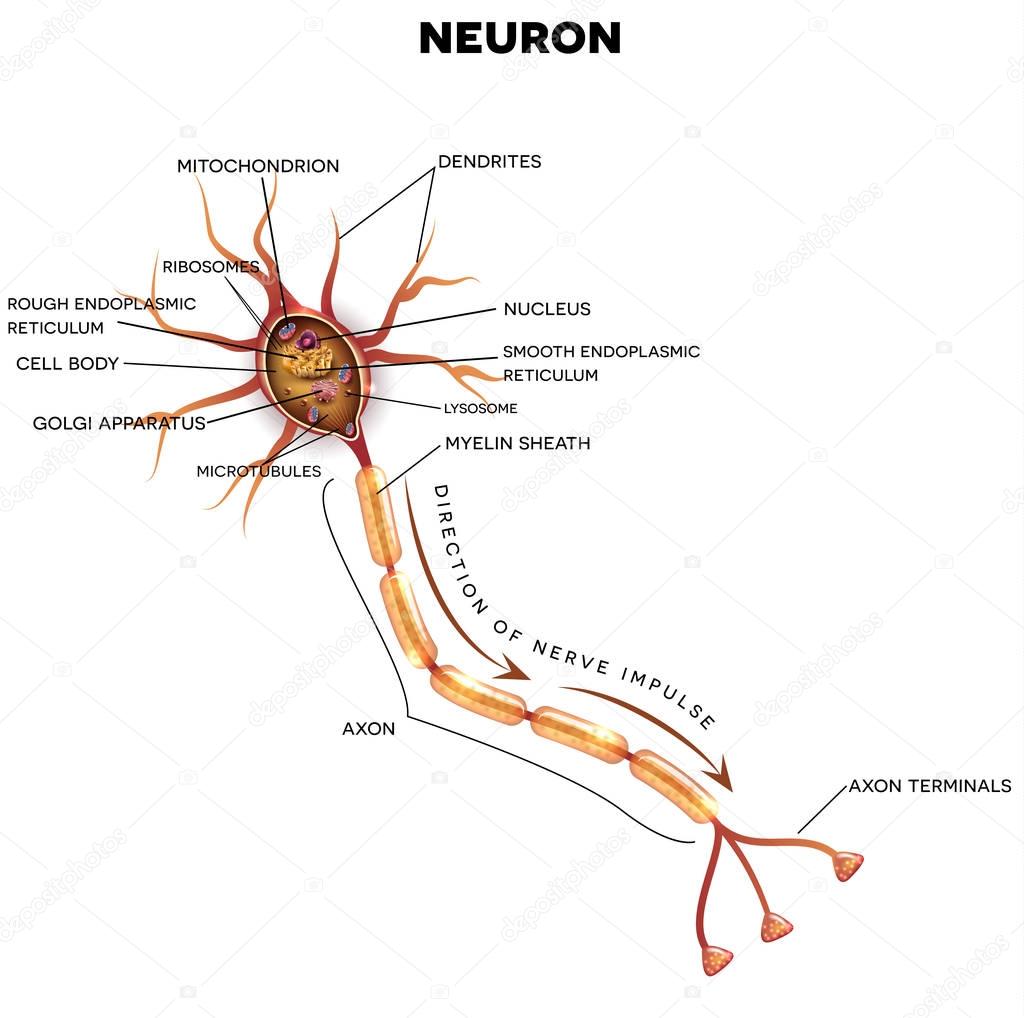
AboutTranscript. Neurons (or nerve cells) are specialized cells that transmit and receive electrical signals in the body. Neurons are composed of three main parts: dendrites, a cell body, and an axon. Signals are received through the dendrites, travel to the cell body, and continue down the axon until they reach the synapse (the communication.
A Junction at Which a Neuron Meets Another Cell
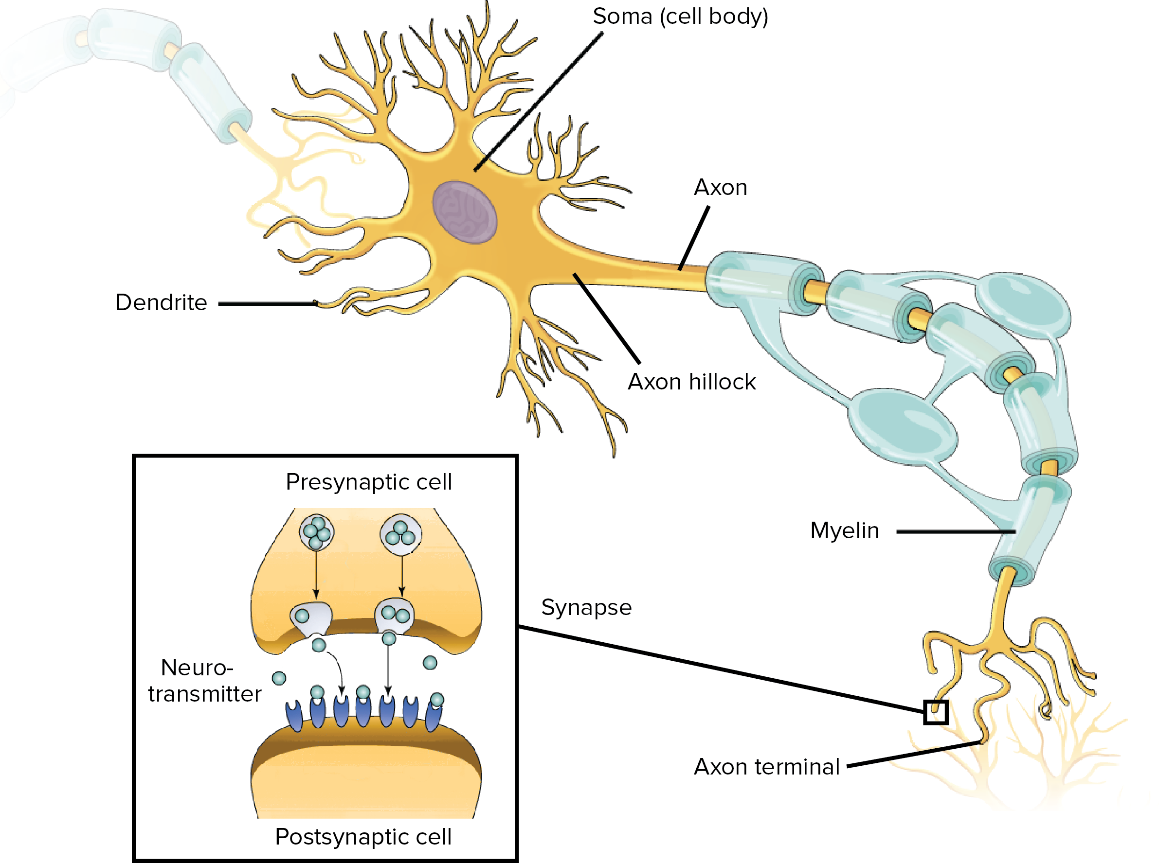
At a synapse, one neuron sends a message to a target neuron—another cell. Most synapses are chemical; these synapses communicate using chemical messengers. Other synapses are electrical; in these synapses, ions flow directly between cells. At a chemical synapse, an action potential triggers the presynaptic neuron to release neurotransmitters.

Nervous Tissue Mediates Perception and Response Anatomy and Physiology I
Neurons are connected to other neurons at synapses and connected to effector organs or cells at neuroeffector junctions. A typical multipolar neuron is comprised of soma or cell body, an axon, and dendrites. The axon is thought of as the part transmitting efferent signals, while the dendrites are receiving afferent signals from their surroundings.

Okay, let’s introduce the brain Exact Approximations
Cerebellum - molecular, Purkinje, granular layers. Peripheral nerves - epineurium, perineurium, endoneurium. This article will explain the histology of neurons, providing you with information about their structure, types, and clinical relevance. It will also cover briefly the histological layers of the central and peripheral nervous systems.
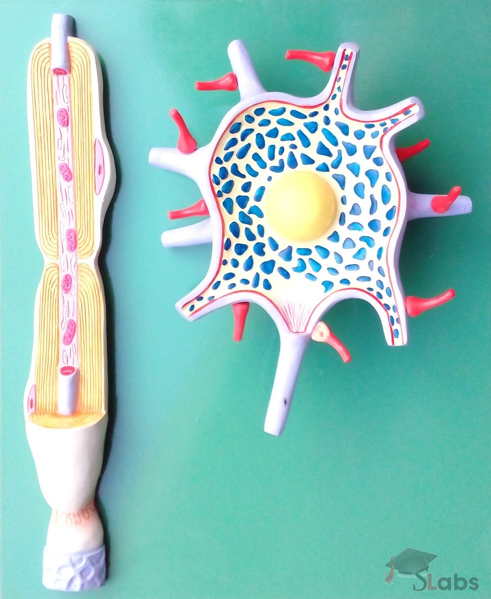
Neuron Model Scholars Labs
A neuron is a nerve cell that processes and transmits information through electrical and chemical signals in the nervous system. Neurons consist of a cell body, dendrites (which receive signals), and an axon (which sends signals). Synaptic connections allow communication between neurons, facilitating the relay of information throughout the body.

12.4 Communication Between Neurons Anatomy & Physiology
The neuron (or nerve cell) is the functional unit of both the central nervous system (CNS) and the peripheral nervous system (PNS). The basic functions of neurons can be summarized into three main tasks: receiving signals, integrating these signals and transmitting the signals to target cells and organs.
Structure of a Neuron Owlcation
Introduction The human brain is perhaps the most complex of all biological systems, with the mature brain composed of more than 100 billion information-processing cells called neurons. [1] The brain is an organ composed of nervous tissue that commands task-evoked responses, movement, senses, emotions, language, communication, thinking, and memory.
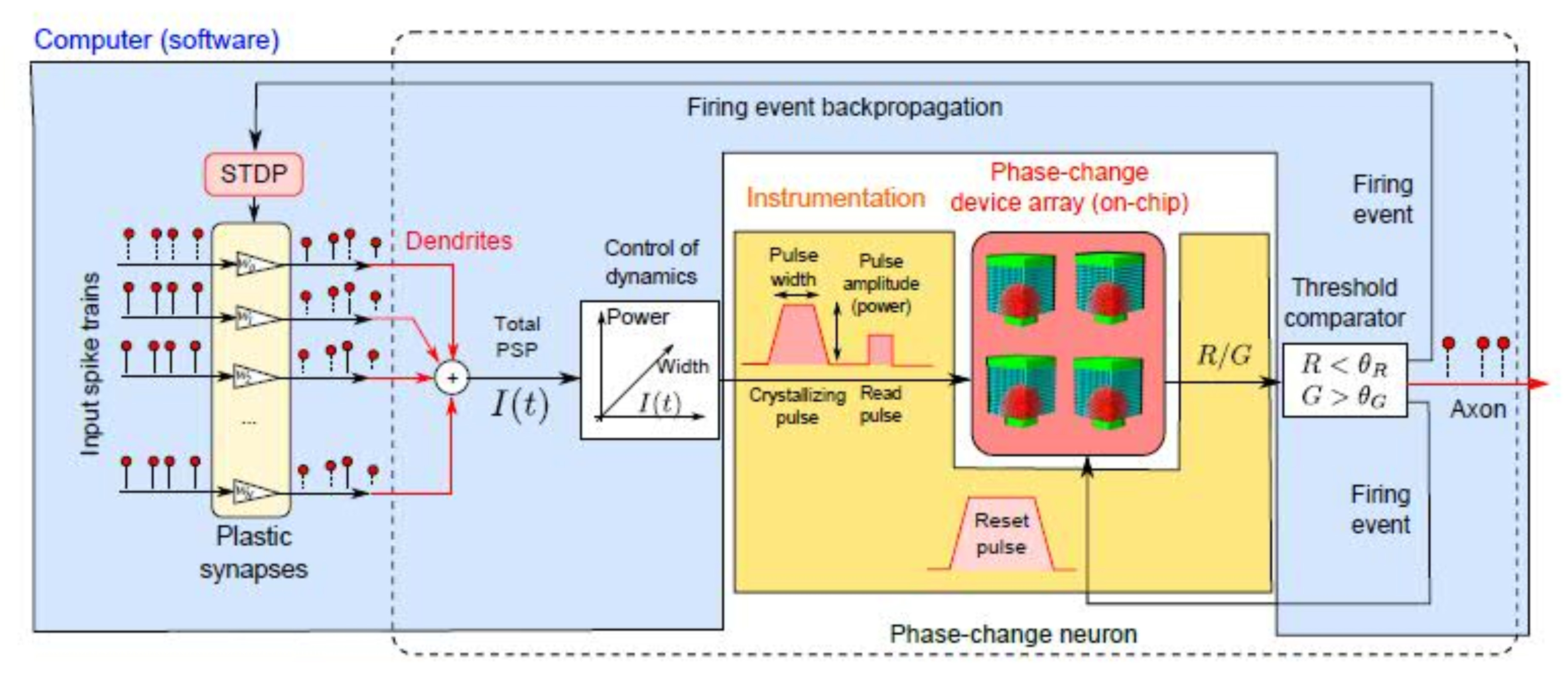
IBM creates world’s first artificial phasechange neurons Ars Technica
The amino acid neurotransmitters glutamate, GABA (γ-aminobutyric acid), and glycine. All of these are amino acids, though GABA is not an amino acid that's found in proteins. Glycine, glutamic acid, and GABA structures. All are amino acids. The biogenic amines dopamine, norepinephrine, epinephrine, serotonin, and histamine, which are made from.
Neuron, Nervenzelle Anatomie — Stockvektor © megija 153364746
How Do Nerve Signals Work? Nerve signals actually come down to some interesting chemistry. Nerve cells communicate with each other using chemicals called neurotransmitters. If the combination of neurotransmitters is correct, then they can cause an electrical current to sweep down the nerve cell.

Neuronal Cell Anatomy Illustration
Biology Biology Article Diagram Of Neuron Diagram Of Neuron A neuron is a specialized cell, primarily involved in transmitting information through electrical and chemical signals. They are found in the brain, spinal cord and the peripheral nerves. A neuron is also known as the nerve cell.

Anatomy of the Nervous System Microbiology
There are three main types of neurons: Motor neurons make the connection between the brain and muscles throughout the body. These neurons transmit electrical impulses containing information to skeletal muscles and smooth muscles. Motor neurons control all of our body movement. Sensory neurons are neurons that let us feel sensation.
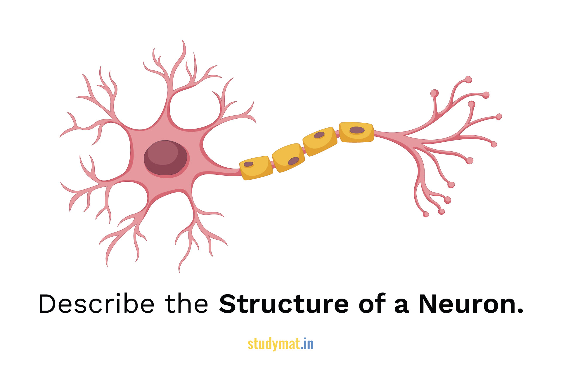
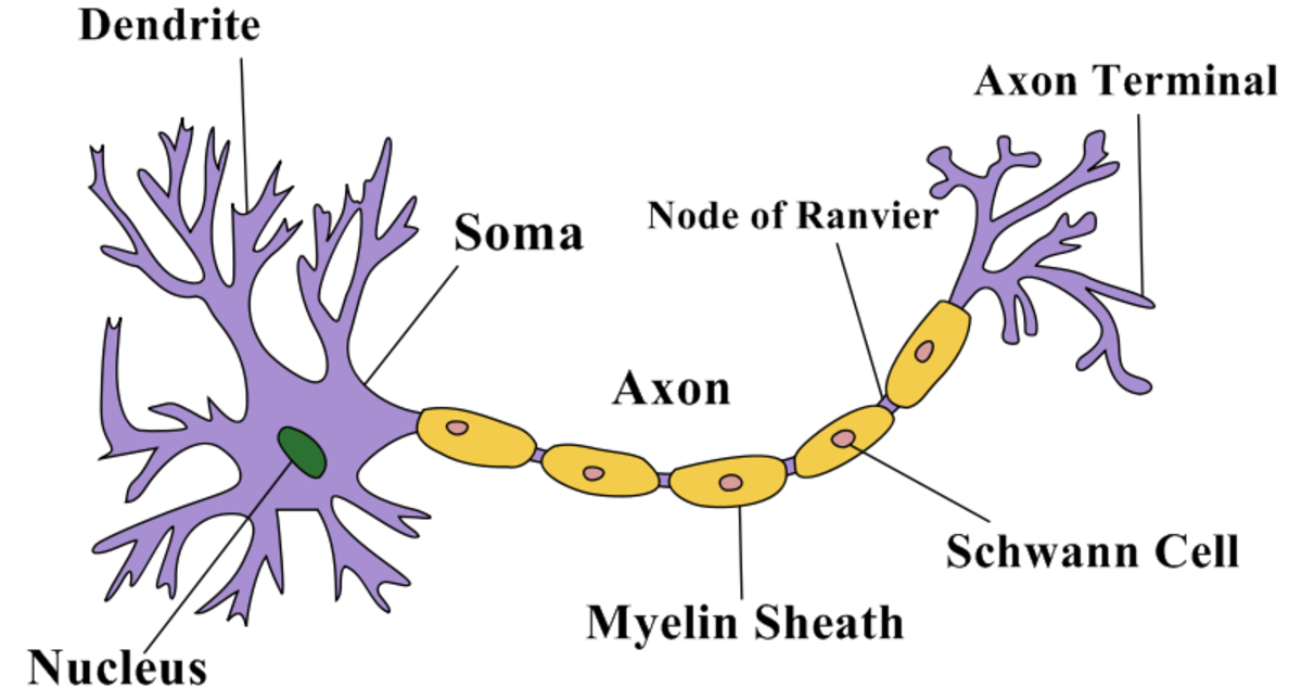
Structure of a Neuron. STUDYMAT
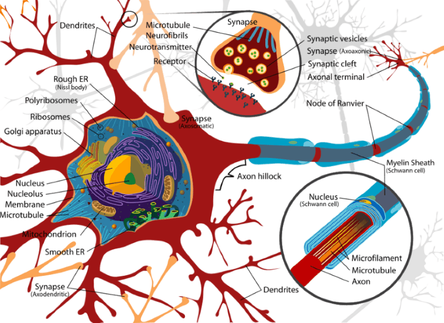
Parts of an Axon. a) Axon hillock - The part of the axon which remains attached to the cell body or soma. b) Myelin sheath - The layer of fatty acid produced from specialized cells called Schwann cells that are wrapped around the axon. c) Nodes of Ranvier - The gaps between the discontinuous myelin sheath that is running along the axon.

Draw A Neuron And Label Its Parts Q10 A Draw The Structure Of Neuron And Label Cell Body And
An Easy Guide to Neuron Anatomy with Diagrams Anatomy Types Function Research Takeaway Neurons, also known as nerve cells, send and receive signals from your brain. While neurons have a lot.

Well Labelled Diagram Of Neuron Porn Sex Picture
of the neuron another general structure of the neuron cell body (soma) A B FIGURE 1-1A and B Generic structure of neuron. This is an artist'sconception of the generic structure of a neuron. All neurons have a cell body known as the soma, which is the command center of the nerve and contains the nucleus of the cell.
Definitions of Human Brain Components Disabled World
Learn about the neuron diagram, structure, and function. Discover and discuss the meaning of dendrites, axon, membrane polarization, membrane depolarization, and synapses. Updated: 11/21/2023.

तंत्रिका की संरचना और कार्य \ Neuron structure and function in Hindi \ Science cbse and ncert
About Transcript This video introduces the structure and structural types of neurons. Explore the structure of neurons, their types, and functions. Uncover the roles of dendrites, axons, and the soma. Learn about the axon hillock, axon terminals, and the myelin sheath.